Services
Thoracoscopy

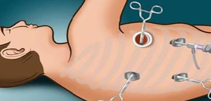
A thoracoscopy is a common procedure to look at the surface of your lungs and the area around your lungs (pleural space). Your surgeon uses a thoracoscope (a thin camera with a light) to see these areas. They can see your diaphragm, esophagus, chest wall and other areas as well.
Your surgeon can use thoracoscopy as part of video-assisted thoracoscopic surgery (VATS), a minimally invasive chest surgery and do certain surgical procedures without the need for opening up the chest. They can project images to a video monitor in the operating room while they work.
What type of procedure is a thoracoscopy?
Thoracoscopy is diagnostic when it’s used to look in your chest or take samples (biopsies) of tissue.
Therapeutic thoracoscopy is used as part of minimally invasive surgery to treat a specific problem.
When is a thoracoscopy used?
Your surgeon can use a thoracoscopy or video-assisted thoracoscopy surgery when they need to:
- Get information they couldn’t get from a chest X-ray, CT scan or ultrasound.
- Remove some of your pleura (the inner layer of the chest wall).
- Remove damaged parts of your lung (lung volume reduction surgery).
- Take air pockets out of your lungs, and remove the damaged area causing air leak.
- Remove a diseased part of your lung (lung resection).
- Drain extra fluid from your pleural space and use medicine to keep fluid from collecting again (pleurodesis).
- Do advanced reconstruction surgery in case of esophageal atresia (partially/incompletely developed food pipe) and fistulas (connection between food pipe and air pipe)
- Diaphragmatic hernia repair


